Mass spectrometry analysis of saponins

Saponins are amphiphilic molecules of pharmaceutical interest and most of their biological activities (i.e., cytotoxic, hemolytic, fungicide, etc.) are associated to their membranolytic properties. These molecules are secondary metabolites present in numerous plants and in some marine animals, such as sea cucumbers and starfishes. Structurally, all saponins correspond to the combination of a hydrophilic glycan, consisting of sugar chain(s), linked to a hydrophobic triterpenoidic or steroidic aglycone, named the sapogenin. Saponins present a high structural diversity and their structural characterization remains extremely challenging. Ideally, saponin structures are best established using nuclear magnetic resonance experiments conducted on isolated molecules. However, the extreme structural diversity of saponins makes them ch
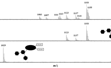
allenging from a structural analysis point of view since, most of the time, saponin extracts consist in a huge number of congeners presenting only subtle structural differences. In the present review, we wish to offer an overview of the literature related to the development of mass spectrometry for the study of saponins. This review will demonstrate that most of the past and current mass spectrometry methods, including electron, electrospray and matrix-assisted laser desorption/ionization ionizations, gas/liquid chromatography coupled to (tandem) mass spectrometry, collision-induced dissociation including MS3 experiments, multiple reaction monitoring based quantification, ion mobility experiments, and so forth, have been used for saponin investigations with great success on enriched extracts but also directly on tissues using imaging methods.